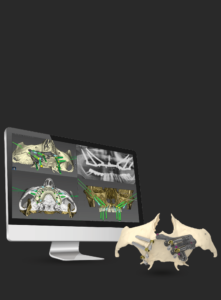

Figure 1A: Initial computed tomography (3D)
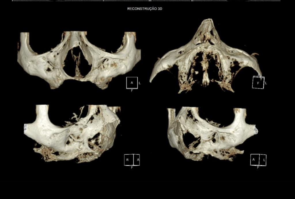
Figure 1B: Initial computed tomography
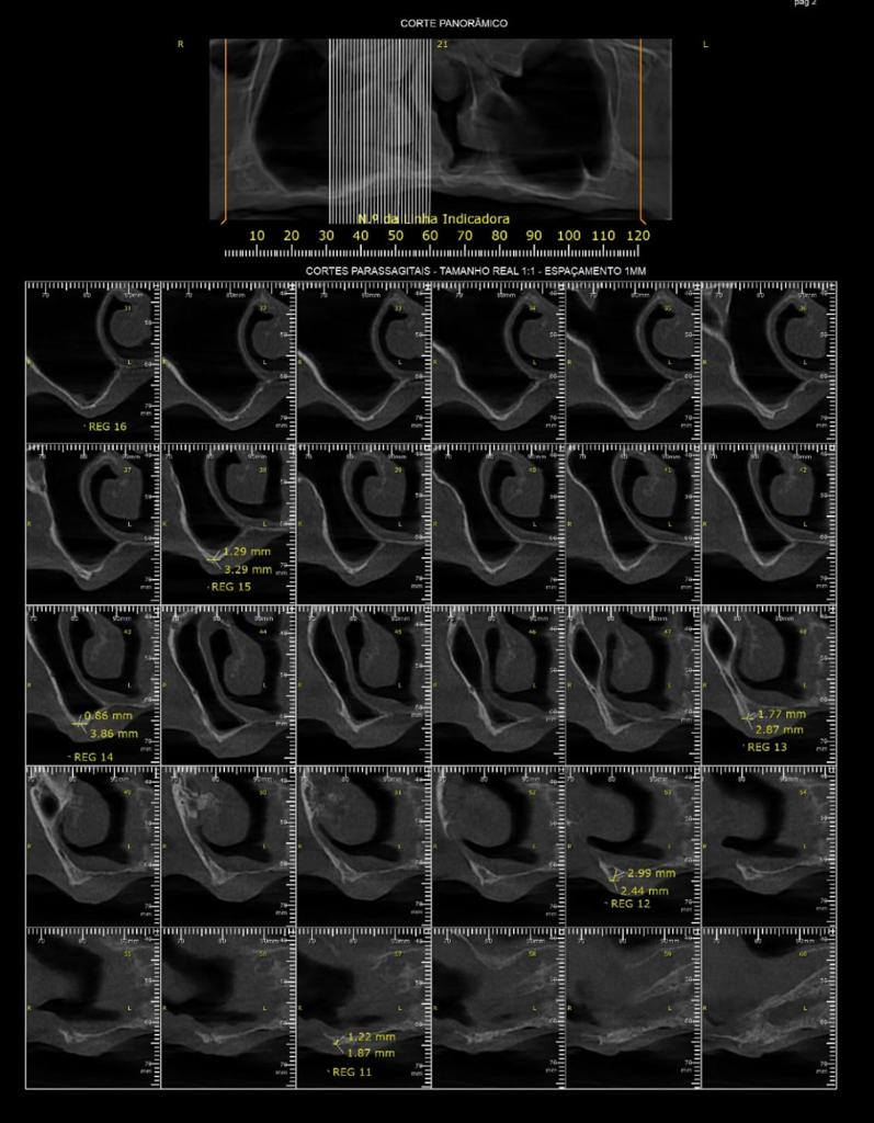
Figure 2: atrophic maxilla (occlusal view)
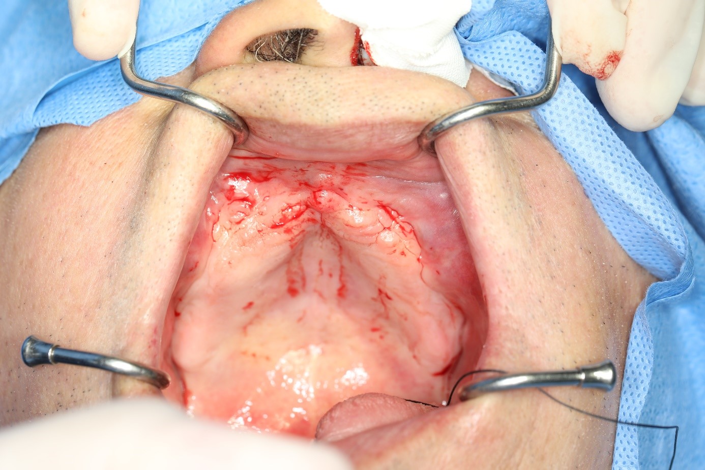
Figure 3: marking of radiographic zones
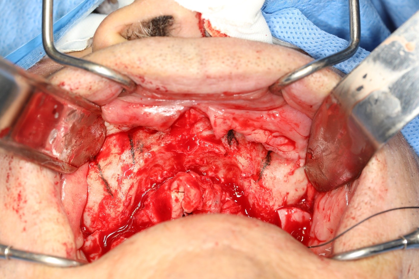
Figure 4: marking of the entry points and paths of the zygomatic implants on the right side
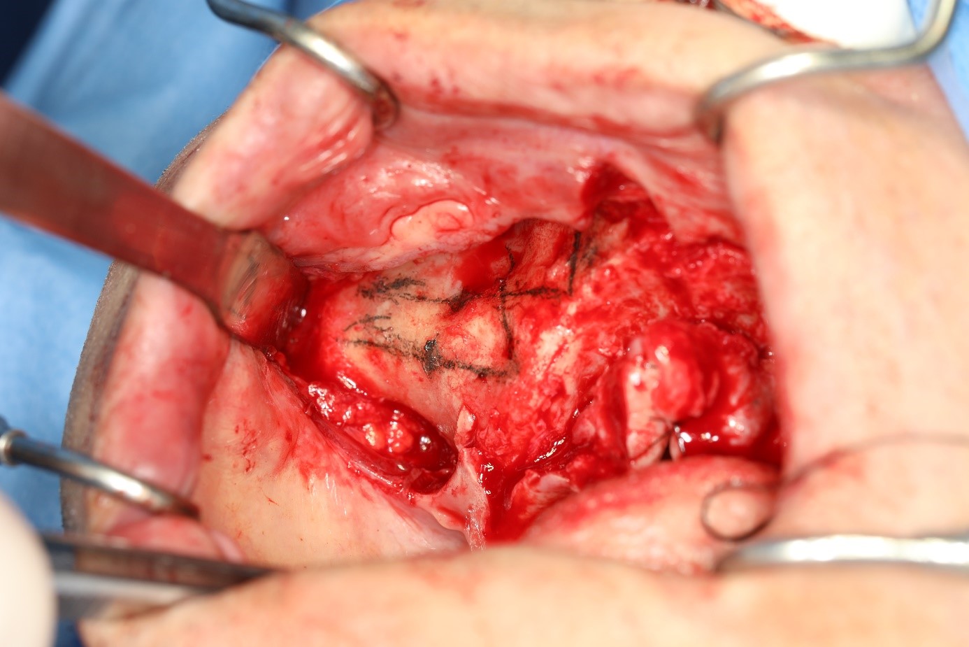
Figure 5: access point performed with the ball drill
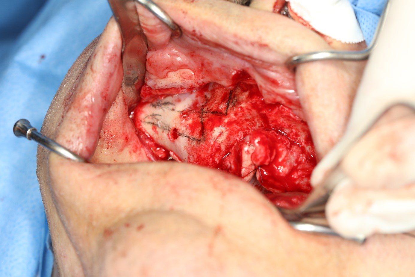
Figure 6: sinus membrane detachment
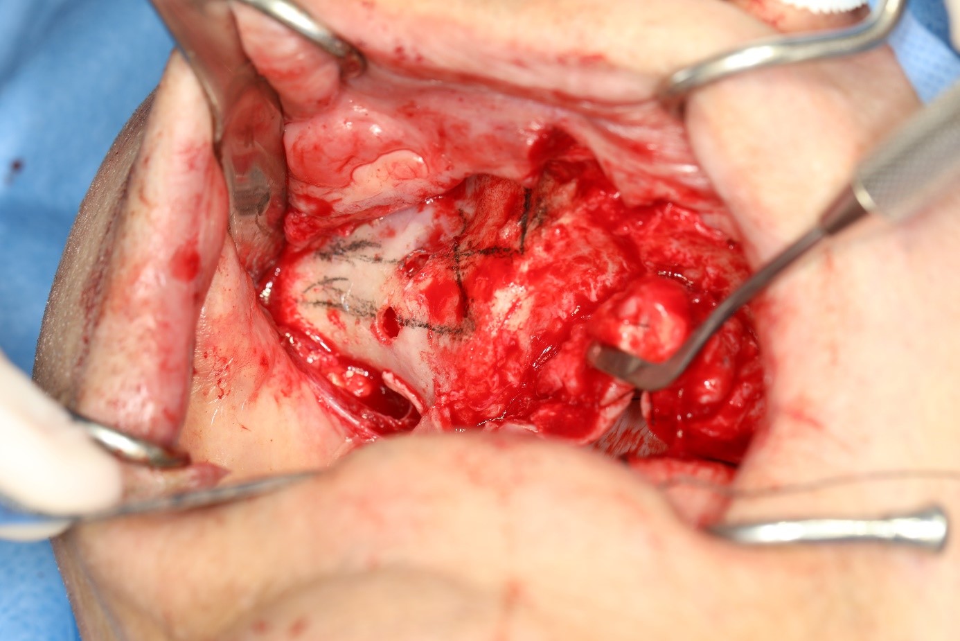
Figure 7: preparation of the 2 channels
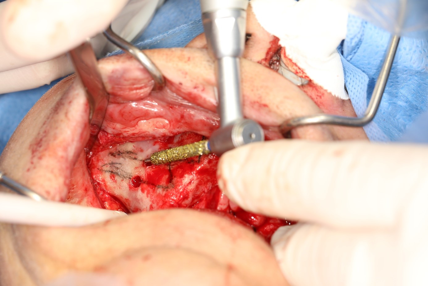
Figure 8: channels made
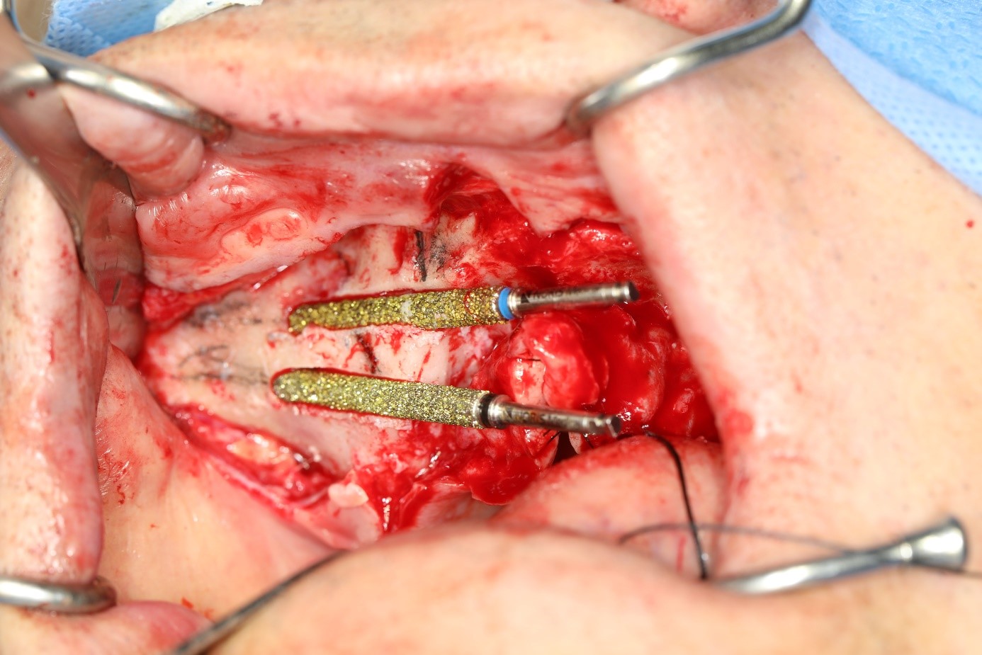
Figure 9: sequence of drills performed (only up to drill number 2)
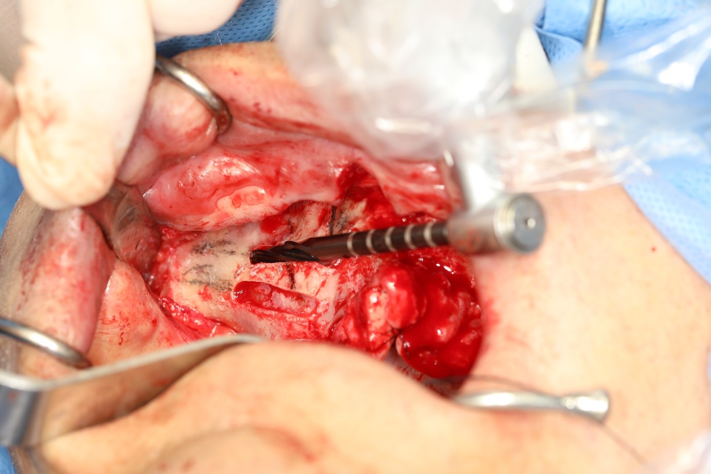
Figure 10: milling performed
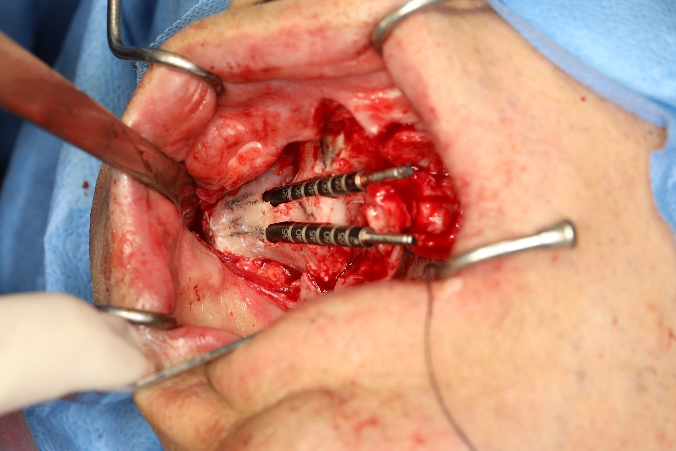
Figure 11: right side zygomatic implants installed
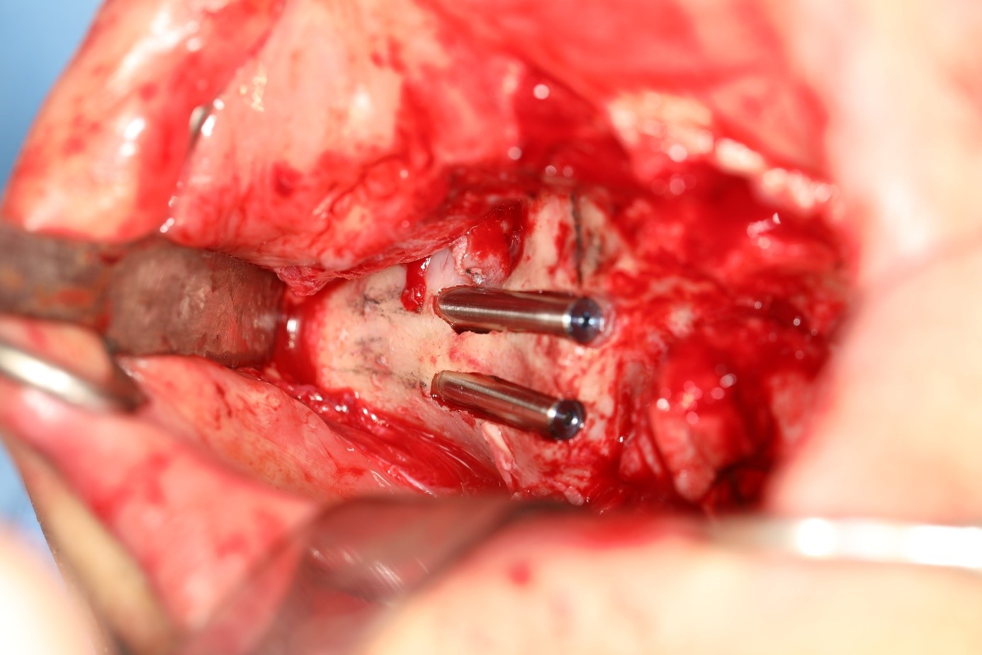
Figure 12: quad zygoma
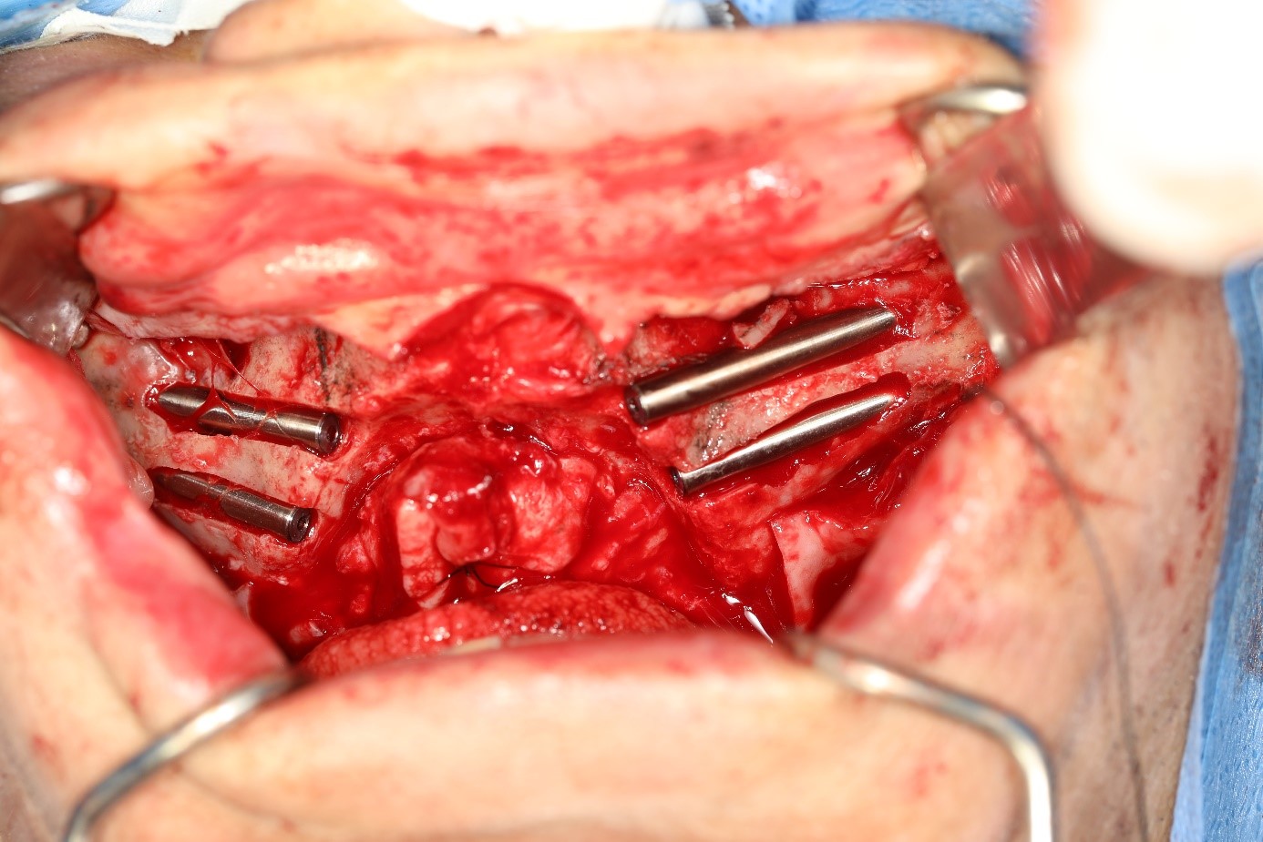
Figure 13: transnasal implant installed
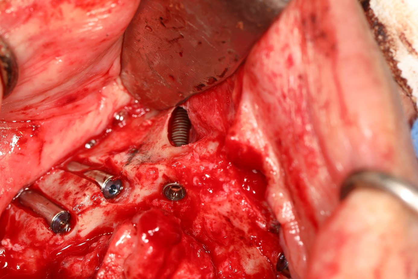
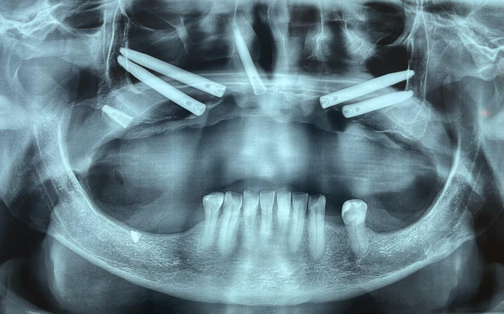
Figure 14: final panoramic radiograph