Figure 1: Initial panoramic X-Ray
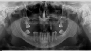
Figure 2A: CT scan anterior region showing bone for conventional implant placement in the region of tooth 22.
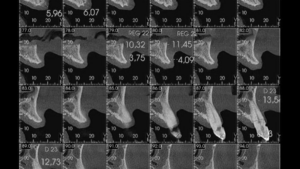
Figure 2B: CT scan anterior region showing bone for conventional implant placement in the region of tooth 12.
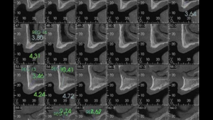
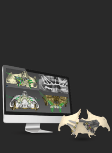
Figure 2C: Computed Tomography (3D Image)
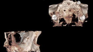
Figure 3: Regularisation of the bone and marking of radiographic areas
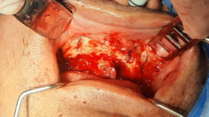
Figure 4: Preparation of the zygomatic implant path (left side)
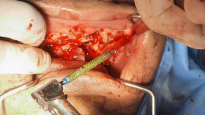
Figure 5: Probe measurement to determine zygomatic length left side
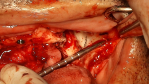
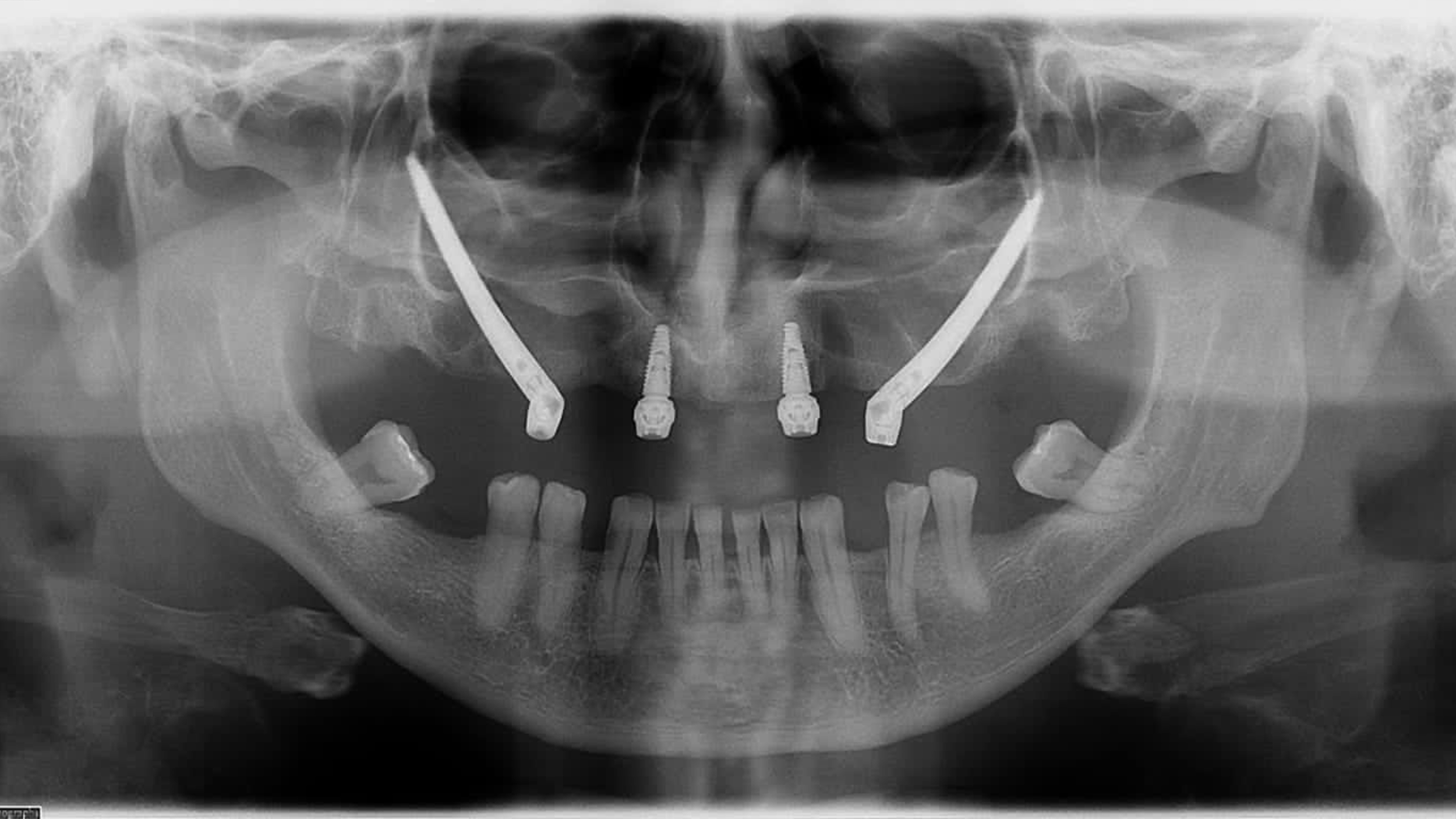
Figure 6: Zygomatic and conventional implants installed
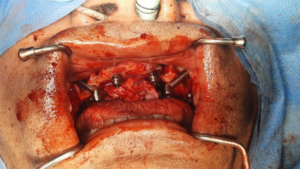
Figure 7: Final panoramic X-ray
